A tiny brain protein may be quietly disarming Alzheimer’s
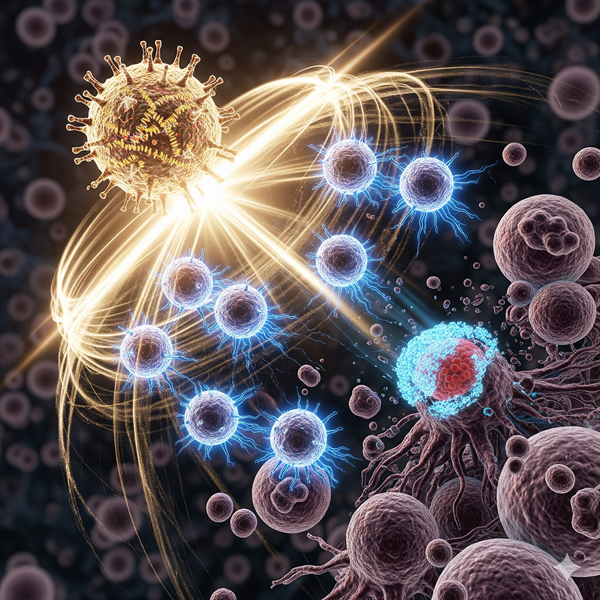
If Alzheimer’s disease is a fire, amyloid‑beta (Aβ) clumps are the sparks that help it spread. A new line of research suggests the brain may already deploy a miniature fire suppressor: a protein called midkine. And if early results hold, learning how to boost this natural defense could change how we prevent or slow the disease.
what’s new here
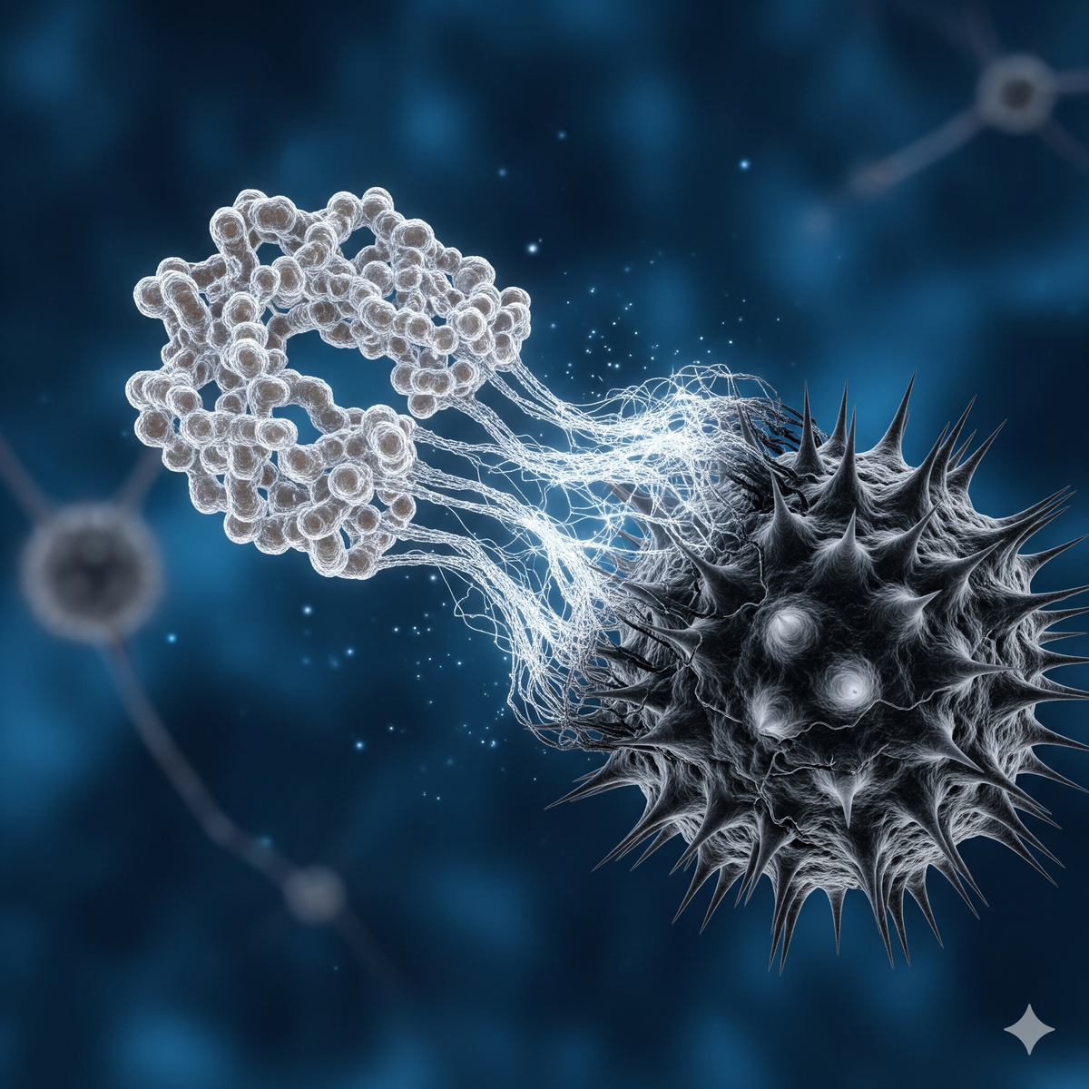
Researchers led by St. Jude Children’s Research Hospital report that midkine can bind directly to Aβ and keep it from assembling into the long, sticky fibrils that harden into plaques. In lab assays, purified midkine slowed fibril formation of both Aβ40 and Aβ42 and kept Aβ in a less structured state. Imaging revealed fewer classical fibrils when midkine was present, and solution NMR showed Aβ signals persisted longer, consistent with less aggregation. Kinetic modeling suggested midkine dampens key steps in the assembly process, including secondary nucleation and elongation (Res Square preprint, NIH‑funded) (https://pmc.ncbi.nlm.nih.gov/articles/PMC11177971/).
The team then asked the crucial question: does this matter in a living brain? In an aggressive amyloidosis mouse model (5xFAD), knocking out the midkine gene caused more insoluble Aβ, denser plaques, and heightened microglial activation. Unbiased proteomics showed broader accumulation of Aβ‑correlated proteins when midkine was missing, pointing to a systemwide loss of protection (preprint above).
These findings build on peer‑reviewed work from 2011 showing that midkine binds Aβ with mid‑nanomolar affinity, with its C‑terminal half grabbing Aβ more tightly than the N‑terminal half. Heparin competed with Aβ for midkine’s binding site, hinting at how the interaction is organized. Importantly, aged midkine‑knockout mice accumulated more spontaneous Aβ deposits than wild‑type littermates, and midkine promoted microglial migration in vitro, suggesting a second protective role in cleanup (International Archives of Medicine, 2011) (https://intarchmed.biomedcentral.com/articles/10.1186/1755-7682-4-1). Earlier cell studies had already hinted at these effects, with midkine reducing Aβ fibrillation and toxicity in neuronal cells (https://pubmed.ncbi.nlm.nih.gov/10214973/).
In short: multiple lines of evidence point to midkine as a built‑in resilience factor against Aβ pathology.
quick primer: key terms in plain language
- Amyloid‑beta (Aβ): a small protein fragment that can stick to itself; over time, these clumps can injure brain cells.
- Fibrils and plaques: fibrils are long fibers formed by stacked Aβ; plaques are dense deposits of fibrils in brain tissue.
- Secondary nucleation and elongation: microscopic stages in which existing Aβ seeds create more aggregates (secondary nucleation) and fibrils grow longer (elongation).
- Microglia: the brain’s resident immune cells that can help clear debris, including Aβ.
why this matters
- A shift from demolition to prevention: Current anti‑Aβ antibodies like lecanemab work largely by marking plaques for removal and have shown modest clinical benefit (NEJM, 2023) (https://www.nejm.org/doi/full/10.1056/NEJMoa2212948). If midkine prevents clumps from forming in the first place, it could complement or, in some cases, precede antibody treatment.
- Biomarker potential: Because midkine levels rise alongside Aβ in human brain proteomes, carefully measuring midkine in cerebrospinal fluid or blood might help track the brain’s own protective response (preprint: https://pmc.ncbi.nlm.nih.gov/articles/PMC11177971/).
- Therapeutic angles: Strategies could include delivering midkine itself, designing peptides or small molecules that mimic its Aβ‑binding surface, or boosting the brain’s midkine production—while rigorously testing safety.
what we still need to learn
- Causality in humans: Midkine is elevated in Alzheimer’s brains—compensation or contributor? The mouse data support a protective role, but human studies must confirm direction and magnitude (preprint: above).
- Safety and specificity: Midkine is a multifunctional growth factor; any therapy must avoid unintended effects in other tissues. Dose, timing, and brain delivery routes will be critical endpoints in preclinical development.
- Clinical synergy: Could midkine‑based approaches pair with existing antibodies (e.g., lecanemab) to block new seeds while clearing old deposits? Prospective combination studies would be informative.
a closer look at the experiments
- Binding and structure: Surface plasmon resonance quantified a midkine–Aβ dissociation constant around 160 nM; domain mapping placed most of the Aβ affinity in midkine’s C‑terminal half, and heparin competed for this interface (2011 study: https://intarchmed.biomedcentral.com/articles/10.1186/1755-7682-4-1).
- Aggregation kinetics: Thioflavin T assays showed slower Aβ40/42 fibrillation with midkine present; circular dichroism indicated less β‑sheet content; electron microscopy visualized fewer mature fibrils; NMR revealed preserved monomeric Aβ signals over time. Kinetic fits via AmyloFit implicated suppression of secondary nucleation (k2) and elongation (k+) (preprint: https://pmc.ncbi.nlm.nih.gov/articles/PMC11177971/).
- In vivo relevance: Removing midkine in 5xFAD mice increased plaque load, insoluble Aβ, and microglial activation, while proteomics captured a broader escalation of amyloid‑linked proteins—evidence that endogenous midkine restrains pathology (preprint: above).
questions to ponder
- If the brain already upregulates midkine near plaques, how can we amplify this response without disrupting other midkine‑dependent processes?
- Would early, preventive midkine‑based therapies help those at genetic risk before symptoms emerge?
- Could midkine levels serve as an early‑warning biomarker of the brain’s battle against Aβ, guiding when to start or pause treatment?
what to watch next
- Peer review and replication of the 2024 preprint results in independent labs.
- Structural studies that map the exact Aβ–midkine contact surfaces to guide drug design.
- Head‑to‑head tests comparing midkine or midkine‑mimetics with approved antibodies, and combination regimens in animal models.
- Safety studies on chronic midkine modulation, including off‑target effects and dosing windows.
conclusion
Alzheimer’s disease affects tens of millions of people worldwide and remains one of our most urgent health challenges (WHO factsheet: https://www.who.int/news-room/fact-sheets/detail/dementia). The emerging picture of midkine as a natural brake on Aβ aggregation offers a fresh, potentially complementary strategy: stop new sparks from igniting while current therapies douse the existing flames. If validated in humans, midkine‑based diagnostics and treatments could help delay onset, slow progression, and reduce the burden on patients and caregivers alike. The next step is clear: rigorous, collaborative research to translate this protective biology into safe, effective tools. What would it mean if the brain’s own tiny guardian became a cornerstone of Alzheimer’s prevention?
Source URL: https://pmc.ncbi.nlm.nih.gov/articles/PMC11177971/